
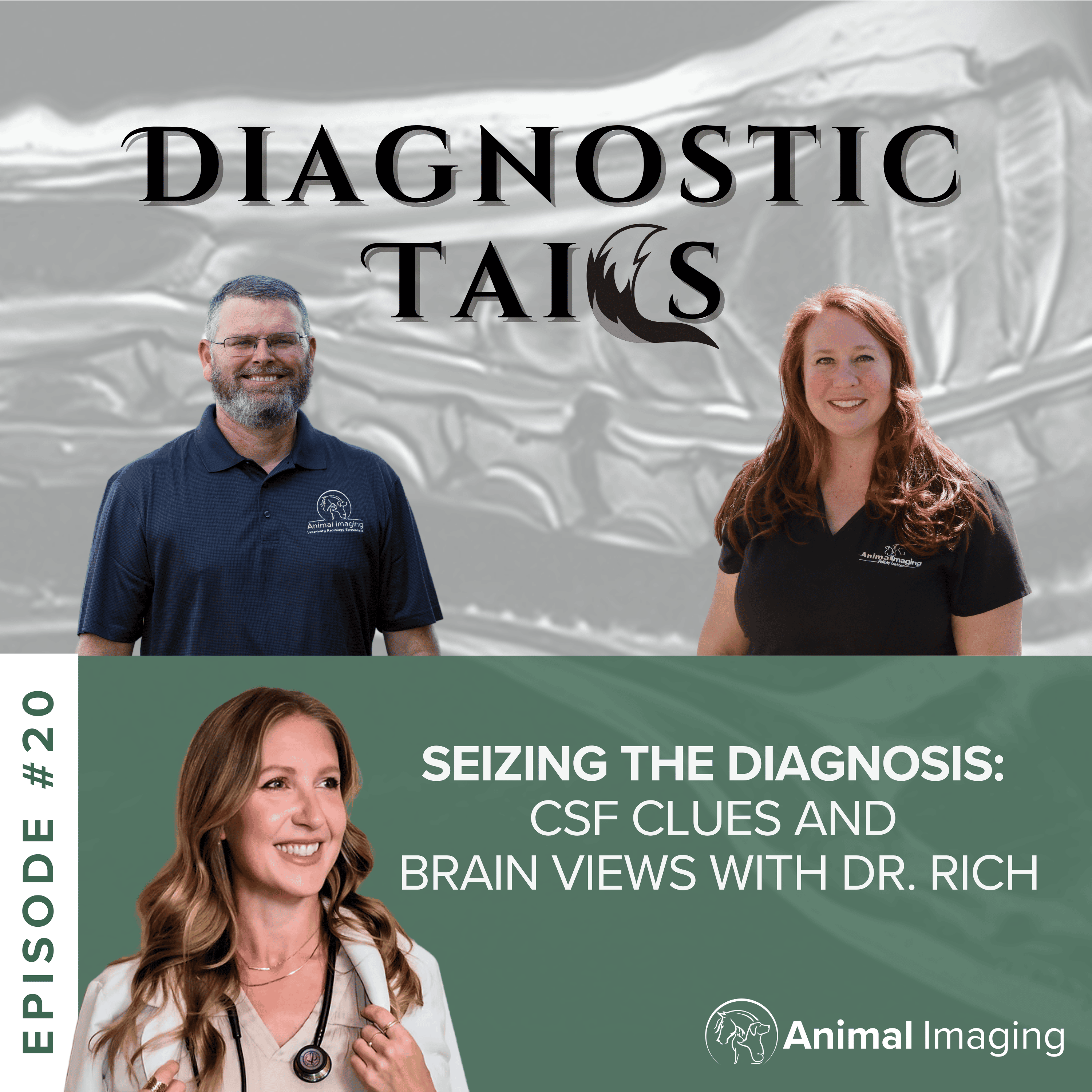
Episode #20
Seizing the Diagnosis: CSF Clues and Brain Views with Dr. Rich
In this episode of Diagnostic Tails , hosts Dr. Amy Armentrout and Dr. Lon Hays are joined by board-certified veterinary neurologist Dr. Jennifer Rich to explore how advanced imaging and cerebrospinal fluid (CSF) analysis work together to diagnose complex neurologic diseases. Through real clinical cases, Dr. Rich explains why an MRI often provides only part of the picture and how a spinal tap can uncover inflammation, infection, and other microscopic changes that imaging alone cannot detect. She also shares a remarkable case in which MRI and CSF analysis identified a treatable middle ear infection that had spread to the brain - saving a dog that otherwise may have been mistakenly treated for a brain tumor. The conversation also covers seizure workups, neurologic localization, when advanced imaging is warranted, and how collaboration between general practitioners, neurologists, radiologists, and pet owners leads to better outcomes for patients. This episode is designed for veterinarians who want a practical approach to neurologic diagnostics and for anyone interested in understanding how advanced imaging can change the course of treatment in patients with neurologic disease. You'll Learn: Why MRI and CSF analysis provide complementary diagnostic information When a spinal tap is recommended during a neurologic workup How MRI can distinguish treatable diseases from life-threatening conditions When seizures warrant advanced imaging and referral The basics of neurologic localization in general practice How to differentiate seizures from other neurologic events Why collaboration between veterinarians and specialists improves patient care How concierge neurology is creating new options for veterinarians and pet owners Timestamps (audio): 00:00 - MRI and Spinal Tap Changed Everything 00:18 - Podcast Welcome and Hosts 01:26 - Meet Neurologist Dr. Jennifer Rich 02:28 - Why Add a Spinal Tap 04:30 - Safety and Timing Tips 06:56 - Dry Taps and Technique 08:05 - Case Otogenic Meningitis 12:48 - Treatable Neuro Conditions 14:21 - Seizure Workup Guidelines 19:25 - Consults and VIP Neurology 23:34 - How to Contact Dr Rich 25:15 - Localizing Lesions for Imaging 27:05 - Neurolocalization Basics 30:19 - Reflexes vs Pain Feeling 32:13 - Movement Disorder Not Seizures 35:51 - When Seizures Need ER 37:12 - Clinic Banter Break 38:18 - Raising Service Dog Floyd 42:20 - Teamwork and Burnout 45:12 - Wrap Up and Resources Key Takeaways: MRI and CSF analysis work together to provide a more complete neurologic diagnosis, revealing both structural and cellular disease. Performing a spinal tap during the initial MRI can avoid a second anesthetic event while providing valuable diagnostic information. Advanced imaging can distinguish treatable conditions from diseases that may appear terminal based on clinical signs alone. Careful neurologic localization and a thorough history help determine the most appropriate imaging study and improve diagnostic efficiency. Not every collapse or convulsive episode is a seizure - recognizing movement disorders and other neurologic conditions leads to more appropriate treatment. Early referral and advanced diagnostics can significantly improve outcomes for patients with neurologic disease. Strong collaboration between general practitioners, imaging specialists, neurologists, and pet owners ensures patients receive the most informed care possible. Connect With Dr. Rich: https://www.virtualvetneurology.com/ Animal Imaging Veterinary Radiology Specialists https://animalimaging.net/ (972) 869-2180 info@animalimaging.net Facebook https://www.facebook.com/AnimalImaging/ Linked In https://www.linkedin.com/company/animalimaging Instagram https://www.instagram.com/animalimaging/?hl=en